Table of Contents
What Is Radiology?
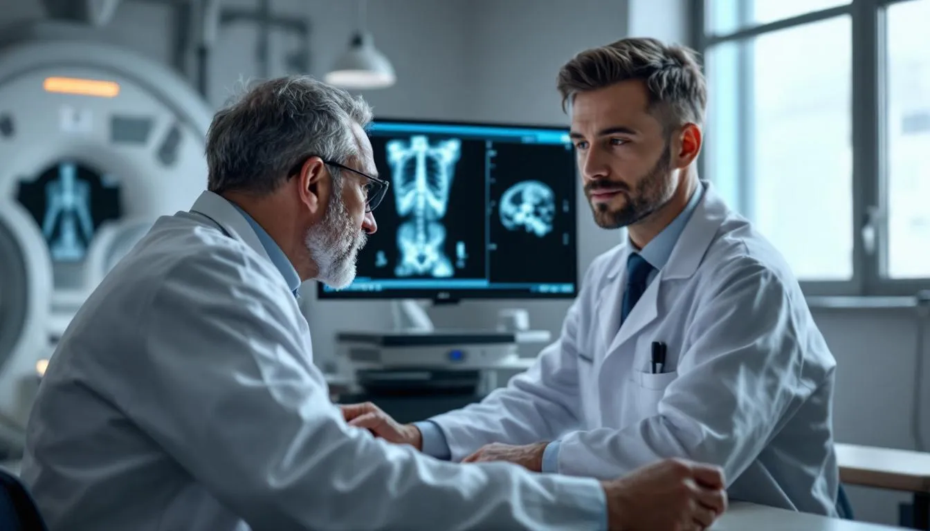
Radiology is the medical specialty that uses imaging technologies — X-rays, CT scans, MRI, ultrasound, and nuclear medicine — to see inside the human body without cutting it open. Radiologists are the physicians who interpret these images, diagnose conditions, and in some cases, perform image-guided treatments. They’re the doctors you rarely meet face-to-face but whose work affects nearly every medical decision involving your body.
Seeing the Invisible
Before radiology existed, doctors could only examine what they could see, touch, or hear through a stethoscope. Internal problems required exploratory surgery — opening someone up just to figure out what was wrong. That changed on November 8, 1895, when German physicist Wilhelm Conrad Roentgen accidentally discovered X-rays while experimenting with cathode ray tubes.
Roentgen noticed that a nearby fluorescent screen glowed even when the tube was shielded. Something was passing through the shield. He spent weeks investigating, eventually producing the first medical X-ray image — his wife Anna Bertha’s hand, complete with wedding ring and visible bones. He called them “X-rays” because he didn’t know what they were. The “X” stood for unknown.
The medical world went wild. Within months, X-ray machines appeared in hospitals across Europe and the United States. For the first time in history, physicians could see bones, detect fractures, and locate foreign objects without surgery. Roentgen won the first Nobel Prize in Physics in 1901.
The early years were also dangerous. Nobody understood radiation’s biological effects. Shoe stores used fluoroscopes to check shoe fit. Early radiologists developed severe radiation burns, and many died of radiation-induced cancers. Marie Curie, who pioneered mobile X-ray units during World War I, likely died from aplastic anemia caused by radiation exposure. These tragedies drove the development of radiation safety standards that protect patients and practitioners today.
The Imaging Toolkit
Modern radiology encompasses several distinct technologies, each with specific strengths.
X-Ray (Radiography)
The oldest and most basic imaging modality. X-rays pass through the body and hit a detector on the other side. Dense structures like bone absorb more X-rays, appearing white on the image. Air (in lungs) absorbs almost none, appearing black. Soft tissues fall somewhere in between.
X-rays are fast, cheap, and widely available. A chest X-ray takes seconds and costs a fraction of what a CT or MRI costs. They’re the first-line test for fractures, pneumonia, heart enlargement, and many other conditions.
Limitations? X-rays compress a three-dimensional body into a two-dimensional image. Structures overlap. Subtle fractures can be invisible. Soft tissue detail is poor. That’s where more advanced modalities come in.
Computed Tomography (CT)
CT scanning — developed in the early 1970s by Godfrey Hounsfield (who won the Nobel Prize for it in 1979) — takes X-rays from multiple angles around the body and uses computer algorithms to reconstruct cross-sectional images. Instead of a flat shadow, you get slices through the body at whatever thickness you choose, typically 0.5-5mm.
Modern CT scanners can image the entire body in under 30 seconds. They’re excellent for detecting tumors, internal bleeding, kidney stones, pulmonary embolism, aortic aneurysms, and complex fractures. CT angiography can visualize blood vessels with injectable contrast dye, often replacing older, more invasive angiography techniques.
The trade-off is radiation dose. A CT scan of the abdomen delivers roughly 100 times the radiation of a chest X-ray. That’s still a small absolute risk for any individual scan, but the cumulative effect of multiple CT scans over a lifetime is a legitimate concern. About 80 million CT scans are performed annually in the U.S. — roughly one for every four people.
Magnetic Resonance Imaging (MRI)
MRI uses strong magnetic fields and radiofrequency pulses to excite hydrogen atoms in your body’s water molecules. When the atoms relax back to their normal state, they emit signals that a computer assembles into images. No ionizing radiation involved.
The result is extraordinary soft tissue contrast. MRI can distinguish between gray matter and white matter in the brain, detect torn ligaments in a knee, show inflammation in organs, and map tumor boundaries with precision that CT can’t match.
The downsides: MRI is slow (20-60 minutes per study), expensive, loud (the machine bangs and clanks as magnetic gradients switch), and claustrophobic (you’re lying inside a narrow tube). Patients with certain metallic implants — pacemakers, cochlear implants, some surgical clips — may not be able to have an MRI safely because the magnet can move or heat metal inside the body. And MRI is terrible at imaging bone cortex and lungs.
Ultrasound
Ultrasound uses high-frequency sound waves. A transducer sends pulses into the body and listens for echoes. Different tissues reflect sound differently, creating a real-time image. No radiation. No magnets. Relatively inexpensive.
Ultrasound is the workhorse of obstetric imaging — most people first encounter it during pregnancy. But it’s also used for evaluating gallbladder stones, blood clots in leg veins (DVT), thyroid nodules, liver disease, and heart function (echocardiography). Newer techniques like shear wave elastography can even measure tissue stiffness, which helps assess liver fibrosis without a biopsy.
The main limitation is that ultrasound can’t penetrate bone or air-filled structures well, making it useless for brain imaging (in adults) and limited for lung evaluation.
Nuclear Medicine
Nuclear medicine flips the script. Instead of sending energy into the body from outside, you inject (or swallow) a tiny amount of radioactive material — a radiotracer — that travels to specific organs or tissues. A special camera detects the gamma rays emitted by the tracer.
PET scans (positron emission tomography) are the best-known nuclear medicine technique. They’re indispensable in oncology — cancer cells metabolize glucose faster than normal cells, so a glucose-based radiotracer (FDG) lights up tumors. PET scans also evaluate brain metabolism in Alzheimer’s disease and heart muscle viability after a heart attack.
Diagnostic vs. Interventional Radiology
Not all radiologists just look at pictures. Interventional radiologists perform minimally invasive procedures guided by imaging — threading catheters through blood vessels, draining abscesses, placing stents, destroying tumors with heat or cold, and stopping bleeding from damaged arteries. It’s surgery without the scalpel.
Some examples of interventional radiology procedures:
Angioplasty and stenting — Opening blocked arteries using a balloon catheter and placing a metal mesh stent to keep it open. All guided by real-time X-ray (fluoroscopy).
Tumor ablation — Inserting a needle into a tumor and destroying it with radiofrequency energy, microwave energy, or cryotherapy (freezing). Used for liver, kidney, and lung tumors.
Embolization — Deliberately blocking blood vessels to stop hemorrhage, shrink uterine fibroids, or cut off blood supply to tumors.
Biopsy — Using CT or ultrasound guidance to insert a needle into a suspicious lesion and extract tissue for pathological analysis. Far less invasive than surgical biopsy.
Interventional radiology has eliminated the need for open surgery in many situations. Uterine fibroid embolization, for instance, treats fibroids through a tiny puncture in the groin artery instead of hysterectomy.
The Radiologist’s Day
A typical diagnostic radiologist reads 50-100 studies per day. Each study might have hundreds of images (a CT scan of the abdomen can generate 500+ individual slices). The radiologist examines every image, dictates a report describing findings and providing a diagnosis or differential diagnosis, and sends it to the ordering physician.
It’s intellectually demanding pattern recognition work. Radiologists are essentially looking for “things that shouldn’t be there” or “things that look different from normal” across thousands of images daily. Miss a small lung nodule on a CT scan, and a cancer might go undetected for months. The pressure is real — radiological misses are a leading source of medical malpractice claims.
AI in Radiology — Hype and Reality
Artificial intelligence has been “about to replace radiologists” since approximately 2016, when Geoffrey Hinton famously said, “We should stop training radiologists now.” It’s 2025, and radiologists are still very much employed.
AI is genuinely good at specific, well-defined tasks: detecting diabetic retinopathy in eye scans, flagging suspicious lung nodules on CT, identifying fractures on X-rays. Several FDA-cleared AI tools assist radiologists by prioritizing urgent cases or providing a “second read.”
But replacing the radiologist? That requires understanding clinical context, integrating information across multiple studies, communicating layered findings, and exercising judgment — things AI still struggles with. The emerging consensus: AI won’t replace radiologists, but radiologists who use AI will replace those who don’t.
Radiation Safety — The Numbers
Every imaging decision involves a benefit-risk calculation. Here are approximate radiation doses for common studies (measured in millisieverts, mSv):
- Chest X-ray: 0.02-0.1 mSv
- Dental X-ray: 0.005 mSv
- Mammogram: 0.4 mSv
- CT head: 2 mSv
- CT abdomen: 8-10 mSv
- CT coronary angiography: 5-15 mSv
For context, average annual background radiation exposure in the U.S. is about 3.1 mSv from natural sources (cosmic rays, radon gas, minerals in the earth). A coast-to-coast flight adds about 0.04 mSv.
The guiding principle in radiology is ALARA — As Low As Reasonably Achievable. Use the minimum radiation necessary to get diagnostic-quality images. Don’t order imaging tests without a clinical reason. Choose non-radiation alternatives (ultrasound, MRI) when they’ll answer the clinical question equally well.
Becoming a Radiologist
The training path: four years of medical school, one year of internship, four years of radiology residency, and often one to two years of fellowship subspecializing in areas like neuroradiology, musculoskeletal radiology, breast imaging, or interventional radiology. That’s 10-11 years of post-college education.
Radiology consistently ranks among the most competitive medical specialties. It also ranks among the highest-paying — median salaries exceed $400,000 in the U.S. The combination of intellectual challenge, technological focus, and quality of life (fewer emergencies than surgery, no overnight call in many practices) makes it a popular career choice for medical students who enjoy visual problem-solving.
Frequently Asked Questions
Is radiation from medical imaging dangerous?
The risk is generally very small but not zero. A single chest X-ray exposes you to about 0.1 mSv of radiation — equivalent to roughly 10 days of natural background radiation. A CT scan delivers more (5-20 mSv depending on the body part), which is why doctors weigh the diagnostic benefit against radiation exposure before ordering one. MRI and ultrasound use no ionizing radiation at all.
What is the difference between a CT scan and an MRI?
CT scans use X-rays and are excellent for bones, bleeding, and lung imaging — they're fast (often under a minute) and widely available. MRI uses magnetic fields and radio waves, producing superior soft tissue detail — ideal for brains, joints, and spinal cords. CT is usually the first choice in emergencies because of speed. MRI is better for planned, detailed soft tissue evaluation.
Why do radiologists work in dark rooms?
Ambient light reduces contrast on monitors, making it harder to spot subtle findings. A bright room can cause you to miss a faint fracture line or a small tumor. Modern radiology monitors are calibrated to specific brightness levels, and dimming the room maximizes the radiologist's ability to detect abnormalities. It's not a style choice — it's diagnostic accuracy.
Can radiology detect cancer?
Yes, imaging is one of the primary tools for detecting cancer. Mammography screens for breast cancer, CT scans detect lung tumors, and MRI evaluates brain and soft tissue cancers. However, imaging alone usually can't confirm cancer — a biopsy (tissue sample examined under a microscope) is typically needed for definitive diagnosis. Imaging tells you something suspicious is there; pathology tells you what it is.
Further Reading
Cite this article
APA
WhatIs.site. (2025). What Is Radiology?. Retrieved May 27, 2026, from https://whatis.site/radiology MLA
"What Is Radiology?." WhatIs.site, July 15, 2025, https://whatis.site/radiology. Accessed May 27, 2026. Chicago
WhatIs.site. "What Is Radiology?." Last modified May 12, 2026. https://whatis.site/radiology. HTML
<a href="https://whatis.site/radiology">What Is Radiology?</a> — WhatIs.site Related Articles
What Is Anatomy?
Anatomy is the study of body structure in living organisms. Learn about gross and microscopic anatomy, organ systems, history, and why it matters in medicine.
scienceWhat Is Biology?
Biology is the scientific study of living organisms and life processes. Learn about cells, genetics, evolution, ecosystems, and the major branches of biology.
scienceWhat Is Cell Biology?
Cell biology is the scientific discipline that studies cells — the basic structural, functional, and biological units of all known living organisms.