Table of Contents
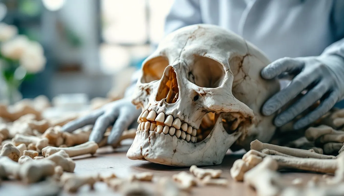
What Is Osteology?
Osteology is the scientific study of bones. That two-word definition covers an enormous amount of ground — from the cellular biology of bone growth to the forensic identification of murder victims, from the evolutionary history written in fossil skeletons to the medical treatment of osteoporosis. Bones are the most durable part of the body, the last thing to decompose, and often the only biological evidence left behind by past lives. Studying them tells us who lived, how they lived, what they ate, how they died, and how they were related to other species.
Bone as Living Tissue
The first thing to understand about bones is that they’re not dead scaffolding. Bones are living, metabolically active organs that constantly remodel themselves throughout life.
What Bones Are Made Of
Bone tissue is a composite material — a combination of organic and inorganic components that together produce a material stronger than steel per unit weight and more flexible than concrete.
The organic component (about 35% by weight) is primarily Type I collagen — long, rope-like protein fibers that provide flexibility and tensile strength. If you remove the mineral component from a bone (by soaking it in acid), the remaining collagen matrix is rubbery and flexible — you can literally tie a bone in a knot.
The inorganic component (about 65% by weight) is hydroxyapatite — a crystalline calcium phosphate mineral that provides hardness and compressive strength. If you burn away the organic component, the remaining mineral is hard but brittle — it shatters like ceramic when dropped.
Together, collagen and hydroxyapatite create a material that resists both bending and compression. This composite principle is the same one used in fiberglass (glass fibers in resin) and reinforced concrete (steel rebar in cement). Biology figured it out about 500 million years ago.
The Cells Inside
Three cell types maintain bone tissue:
Osteoblasts build new bone. They secrete collagen and other proteins that form the organic matrix (osteoid), then mineralize it by depositing hydroxyapatite crystals. When an osteoblast becomes completely surrounded by the bone it has built, it becomes an osteocyte.
Osteocytes are the most abundant bone cells — about 90-95% of all bone cells. They live inside tiny chambers (lacunae) connected by microscopic channels (canaliculi) that form a communication network throughout the bone. Osteocytes sense mechanical loads and orchestrate the remodeling process, telling osteoblasts where to build and osteoclasts where to remove.
Osteoclasts destroy bone. They’re large, multinucleated cells that dissolve both the mineral and organic components. This sounds counterproductive, but it’s essential. Bone remodeling — the continuous cycle of destruction and rebuilding — allows bones to repair microdamage, adapt to changing mechanical loads, and release calcium when the body needs it.
Your entire skeleton is replaced roughly every 7-10 years through this remodeling process. The bones you have now are not the same physical bones you had a decade ago.
Bone Architecture
Bones aren’t solid throughout. They come in two structural forms:
Cortical (compact) bone forms the dense outer shell. Under a microscope, it’s organized into concentric cylinders called osteons (or Haversian systems), each centered on a blood vessel. This architecture maximizes strength while maintaining blood supply.
Trabecular (spongy) bone fills the interior of flat bones, vertebrae, and the ends of long bones. It consists of a lattice of thin struts (trabeculae) oriented along the lines of mechanical stress. This is not random — the trabeculae align themselves to resist the forces the bone actually experiences, a phenomenon first described by Julius Wolff in 1892 (Wolff’s Law). An X-ray of the proximal femur reveals trabecular patterns that match the stress lines predicted by engineering analysis perfectly.
This adaptive architecture means bones literally sculpt themselves to handle the loads placed on them. Tennis players develop thicker cortical bone in their dominant arm. Astronauts lose bone density in space because the absence of gravity removes the mechanical stimulus for bone maintenance. Your bones are a record of the forces your body has experienced.
The Human Skeleton: Structure and Function
The adult human skeleton contains 206 bones, divided into the axial skeleton (80 bones forming the central axis — skull, vertebral column, ribcage) and the appendicular skeleton (126 bones forming the limbs and their attachments — shoulder girdle, arms, hands, pelvic girdle, legs, feet).
The Skull
The skull is actually 22 bones — 8 forming the cranium (braincase) and 14 forming the face. In adults, most cranial bones are fused at suture lines. In infants, gaps between skull bones (fontanelles, or “soft spots”) allow the brain to grow and the skull to compress during birth. The anterior fontanelle doesn’t fully close until about 18 months of age.
The skull protects the brain, houses the sensory organs (eyes, ears, nose), and provides the framework for facial muscles. Its shape varies significantly between populations, ages, and sexes — information that forensic osteologists use for identification.
The Vertebral Column
The spine consists of 33 vertebrae (7 cervical, 12 thoracic, 5 lumbar, 5 sacral fused into the sacrum, and 4 coccygeal fused into the coccyx). Its S-shaped curve — unique among primates — is an adaptation for bipedal locomotion. The curve distributes weight efficiently and absorbs shock during walking and running.
Intervertebral discs between vertebrae are fibrocartilage cushions that allow flexibility while bearing compressive loads. Disc degeneration with age is nearly universal and a leading cause of back pain — affecting about 80% of adults at some point in their lives.
Long Bones
Long bones (femur, tibia, humerus, radius, ulna) are the structural pillars of the limbs. Each has a shaft (diaphysis) of thick cortical bone surrounding a medullary cavity filled with marrow, and two ends (epiphyses) covered with articular cartilage for smooth joint movement.
The femur is the longest and strongest bone in the body. It can withstand compressive forces of about 1,700 pounds before fracturing. Its neck angle (about 125 degrees in adults) is an adaptation for efficient bipedal walking — a feature that physical anthropology) uses to distinguish human from non-human fossils and to track the evolution of upright walking.
The Hand
The human hand contains 27 bones — 8 carpals, 5 metacarpals, and 14 phalanges. This elaborate skeleton provides the dexterity that defines human tool use. The opposable thumb — with its saddle-shaped carpometacarpal joint allowing movement in multiple planes — is one of the anatomical features that enabled human technological achievement.
Forensic Osteology: Bones Tell Stories
When forensic science encounters skeletal remains, osteologists extract a remarkable amount of information.
The Biological Profile
From a complete skeleton, a forensic osteologist can determine:
Sex: Male and female skeletons differ systematically. The pelvis is the most reliable indicator — the female pelvis is wider with a broader subpubic angle (90-100 degrees vs. 50-60 degrees in males) to accommodate childbirth. The skull is also useful: males tend to have more prominent brow ridges, larger mastoid processes, and more squared mandibles. Accuracy exceeds 95% when the pelvis is available.
Age: Different skeletal features change at different life stages. In children and adolescents, dental development and epiphyseal fusion (growth plate closure) provide precise age estimates. The medial clavicular epiphysis is the last to fuse — completing between ages 25-30 — making it useful for distinguishing young adults. In older adults, changes to the pubic symphysis face, auricular surface, and sternal rib ends indicate age, though with less precision.
Stature: Long bone length correlates strongly with height. Regression equations developed from populations with known stature can estimate living height from a single femur within about 3-5 centimeters.
Ancestry: Skeletal features vary between geographic populations due to evolutionary adaptation and genetic drift. Craniometric analysis (skull measurements) can estimate broad geographic ancestry, though this is the most debated aspect of forensic osteology — human variation is clinal (gradual) rather than categorical, and ancestry estimation involves probability, not certainty.
Trauma Analysis
Bones record trauma with remarkable clarity. Forensic osteologists distinguish between:
Antemortem trauma — healed injuries showing bone remodeling (callus formation). A healed fracture indicates the person survived the injury.
Perimortem trauma — injuries occurring at or near the time of death, showing no healing. The fracture pattern in fresh bone differs from dry bone — fresh bone bends and splinters, while dry bone cracks cleanly.
Postmortem damage — breakage occurring after death and decomposition, distinguishable by color differences between the fracture surface and the bone surface.
Specific fracture patterns can indicate the weapon or mechanism: blunt force produces depressed fractures with radiating cracks, sharp force produces linear cut marks with smooth walls, and gunshot wounds produce distinctive beveling patterns that indicate the direction of the projectile.
Identification
Skeletal identification can involve comparison with antemortem medical and dental records (healed fractures, surgical implants, dental work) or, increasingly, DNA extraction from bone. The petrous bone (the densest bone in the body, located in the inner ear) preserves DNA exceptionally well — it’s now the preferred sampling location for ancient DNA extraction, allowing genetic identification of individuals dead for thousands of years.
Archaeological Osteology: Reading the Past
In archaeology, human skeletal remains are primary evidence for understanding past populations.
Health and Disease
Ancient bones record disease with surprising detail. Tuberculosis leaves characteristic lesions on vertebrae (Pott’s disease). Syphilis produces distinctive cranial erosions (caries sicca). Iron deficiency creates porous lesions in the skull roof (porotic hyperostosis) and eye sockets (cribra orbitalia). Scurvy (vitamin C deficiency) causes hemorrhaging visible on bone surfaces.
Growth arrest lines (Harris lines) visible in X-rays of long bones record episodes of illness or malnutrition during childhood — each line represents a period when growth stopped and then resumed. These lines provide a record of childhood stress in populations that left no written history.
The transition from hunting-gathering to agriculture — one of the most significant changes in human history — left clear signatures in skeletal remains. Early farming populations show increased dental cavities (from carbohydrate-rich diets), shorter stature (from nutritional deficiency), more infectious disease (from increased population density and proximity to animals), and more arthritis in specific joints (from repetitive agricultural labor).
Activity and Occupation
Bones remodel in response to mechanical stress, so habitual activities leave their mark. Enthesopathies — roughened areas where muscles and ligaments attach — indicate heavy use of specific muscle groups. Medieval archers develop enlarged left arm bones (from holding the bow) and specific changes to the right shoulder and fingers (from drawing the bowstring). Grain grinders develop arthritic changes in the knees and lower spine from hours of kneeling at grinding stones.
Dental wear can reveal occupational habits. Leather workers who used their teeth to hold strips of hide develop distinctive anterior tooth wear. Pipe smokers create notched grooves in their teeth. Seamstresses who bit thread show wear on specific anterior teeth.
Migration and Population Movement
Stable isotope analysis has revolutionized our understanding of ancient migration. Strontium isotope ratios in tooth enamel reflect the local geology where a person grew up (because strontium substitutes for calcium in enamel during childhood). By comparing tooth enamel isotope ratios with local geological values, researchers can determine whether an individual grew up locally or migrated from elsewhere.
This technique has produced remarkable discoveries. The “Amesbury Archer,” buried near Stonehenge around 2300 BCE with rich grave goods, grew up in the Alps — evidence of long-distance travel in the Bronze Age. Analysis of Roman-era cemeteries in Britain reveals individuals who grew up in North Africa, the Middle East, and Scandinavia — demonstrating the cosmopolitan nature of the Roman Empire through skeletal chemistry.
Comparative and Evolutionary Osteology
Comparing skeletal structures across species reveals evolutionary relationships and adaptations.
The vertebrate skeleton follows a remarkably conserved basic plan. A whale’s flipper, a bat’s wing, a horse’s leg, and a human arm all contain the same bones — humerus, radius, ulna, carpals, metacarpals, phalanges — modified for different functions. This homology was recognized before Darwin and became powerful evidence for common descent.
The fossil record of vertebrate skeletons documents major evolutionary transitions: the emergence of jaws in early fish, the transition from water to land (the famous Tiktaalik fossil shows intermediate fin-limb structures), the development of the mammalian middle ear from reptilian jaw bones, and the evolution of human bipedalism shown in fossils like Australopithecus afarensis (“Lucy”) with her intermediate pelvis and femur anatomy.
Paleoanthropology — the study of human evolution — relies heavily on osteology. The shape of a pelvis, the angle of a femur, the curvature of a spine, the position of the foramen magnum (the hole where the spinal cord enters the skull), and the size of a braincase all contribute to understanding how and when our ancestors began walking upright, using tools, and developing larger brains.
Clinical Osteology: Bones in Medicine
Medical applications of osteology are numerous and growing.
Osteoporosis affects an estimated 200 million people worldwide and causes over 8.9 million fractures annually. The disease involves an imbalance in bone remodeling — osteoclasts remove bone faster than osteoblasts replace it, reducing bone density and increasing fracture risk. Post-menopausal women are most affected because estrogen (which suppresses osteoclast activity) drops sharply at menopause.
Dual-energy X-ray absorptiometry (DEXA) scans measure bone mineral density, providing a T-score that compares an individual’s bone density to a healthy young adult. A T-score below -2.5 indicates osteoporosis. Treatment options include bisphosphonates (which inhibit osteoclasts), denosumab (a monoclonal antibody targeting osteoclast differentiation), and newer anabolic agents that stimulate osteoblast activity.
Fracture healing is a complex process involving inflammation, soft callus formation (fibrocartilage), hard callus formation (woven bone), and remodeling (replacement with lamellar bone). The process typically takes 6-12 weeks for a simple fracture but can take months for complex injuries. Understanding the biology of fracture healing has led to improved surgical techniques, bone grafting materials, and emerging treatments using bone morphogenetic proteins (BMPs) and stem cells.
Bone cancer — both primary (osteosarcoma, Ewing sarcoma) and metastatic (cancer spreading from other organs to bone) — is diagnosed and monitored using imaging, biopsy, and increasingly molecular techniques. The skeleton is the third most common site for cancer metastasis, after lung and liver.
Modern Methods
Contemporary osteological research uses technology that has transformed the field.
Computed tomography (CT) scanning creates 3D reconstructions of bones, revealing internal structure without destructive sampling. Micro-CT scanners achieve resolutions below 10 micrometers, revealing trabecular architecture and cortical porosity in detail impossible to see with the naked eye.
3D surface scanning creates digital models of bones for measurement, comparison, and archiving. Geometric morphometrics — statistical analysis of 3D shape — allows quantitative comparison of skeletal features across individuals and populations, replacing subjective visual assessment with rigorous statistical analysis.
Ancient DNA extraction from bone (especially the petrous bone) has enabled genetic analysis of individuals dead for tens of thousands of years. Svante Paabo’s Nobel Prize-winning work on Neanderthal and Denisovan genomes used DNA extracted from ancient bones, revealing interbreeding between archaic humans and modern Homo sapiens.
Histological analysis — thin-section microscopy of bone — reveals growth patterns, age at death (in both humans and other animals), and pathological changes invisible on the bone’s surface. In non-human animals, lines of arrested growth (LAGs) in bone cross-sections function like tree rings, recording annual growth cycles.
Why Osteology Matters
Bones endure. Long after soft tissues decompose, bones remain — carrying information about identity, health, diet, activity, ancestry, and evolution. Osteology extracts that information using techniques from anatomy, biology, chemistry, physics, and statistics.
Whether the goal is identifying disaster victims, understanding how ancient civilizations lived, tracing human evolution, or treating bone diseases in living patients, osteology provides the knowledge foundation. It’s a field where a single bone fragment can tell you whether a person was male or female, young or old, local or migrant, healthy or diseased, and how they died. Few scientific disciplines pack so much information into such durable evidence.
Key Takeaways
Osteology is the study of bones — their biology, structure, development, diseases, and applications in medicine, forensics, archaeology, and evolutionary research. Bone is a living composite tissue constantly remodeled by osteoblasts, osteocytes, and osteoclasts. The human skeleton’s 206 bones provide structure, protection, and a record of individual life history. Forensic osteologists determine sex, age, stature, ancestry, and cause of death from skeletal remains. Archaeological osteologists reconstruct ancient diet, health, occupation, and migration patterns. Clinical osteology addresses diseases like osteoporosis and bone cancer. Modern techniques including CT scanning, stable isotope analysis, and ancient DNA extraction have dramatically expanded what bones can reveal.
Frequently Asked Questions
How many bones are in the adult human body?
The adult human skeleton contains 206 bones. Babies are born with about 270 bones, but many fuse together during development. The exact number can vary slightly between individuals — some people have extra ribs, extra vertebrae, or small accessory bones called sesamoid bones or Wormian bones in the skull.
What is the difference between osteology and orthopedics?
Osteology is the scientific study of bones — their structure, development, diseases, and role across species. Orthopedics is a medical specialty focused on diagnosing and treating conditions of the musculoskeletal system in living patients. Osteology provides the knowledge foundation; orthopedics applies it clinically.
Can bones reveal a person's age at death?
Yes, with reasonable accuracy. In children and adolescents, tooth development, epiphyseal fusion (growth plate closure), and bone length provide age estimates often accurate to within 1-2 years. In adults, changes to the pubic symphysis, sternal rib ends, and cranial sutures provide estimates, though accuracy decreases with age — typically within 5-10 years for older adults.
How long do bones last after death?
Under favorable conditions (dry caves, arid deserts, alkaline soils), bones can survive for millions of years — dinosaur bones are 65+ million years old. Under unfavorable conditions (acidic soils, tropical forests, waterlogged environments), bones can completely dissolve within decades. Burial in neutral to alkaline soil typically preserves bones for thousands of years.
What can ancient bones tell us about past diets?
Quite a lot. Stable isotope analysis of bone collagen and tooth enamel reveals the proportion of plants vs. meat, marine vs. terrestrial food, and specific types of plants (like maize) in the diet. Trace element analysis can indicate dairy consumption, seafood intake, and even geographic origin. Dental wear patterns and dental caries provide additional dietary information.
Further Reading
Cite this article
APA
WhatIs.site. (2026). What Is Osteology?. Retrieved May 27, 2026, from https://whatis.site/osteology MLA
"What Is Osteology?." WhatIs.site, March 6, 2026, https://whatis.site/osteology. Accessed May 27, 2026. Chicago
WhatIs.site. "What Is Osteology?." Last modified May 12, 2026. https://whatis.site/osteology. HTML
<a href="https://whatis.site/osteology">What Is Osteology?</a> — WhatIs.site Related Articles
What Is Anatomy?
Anatomy is the study of body structure in living organisms. Learn about gross and microscopic anatomy, organ systems, history, and why it matters in medicine.
scienceWhat Is Human Anatomy?
Human anatomy is the scientific study of the body's physical structures — from the macroscopic arrangement of organs and bones down to the microscopic.
scienceWhat Is Archaeology?
Archaeology is the study of human history through physical remains like artifacts, buildings, and bones. Learn about methods, famous discoveries, and careers.
scienceWhat Is Forensic Science?
Forensic science applies scientific methods to criminal and civil investigations, from DNA analysis and fingerprinting to toxicology and digital evidence.
scienceWhat Is Physical Anthropology?
Physical anthropology studies human biology, evolution, and variation. Learn how scientists trace our origins through fossils, genetics, and primatology.