Table of Contents
What Is Ophthalmology?
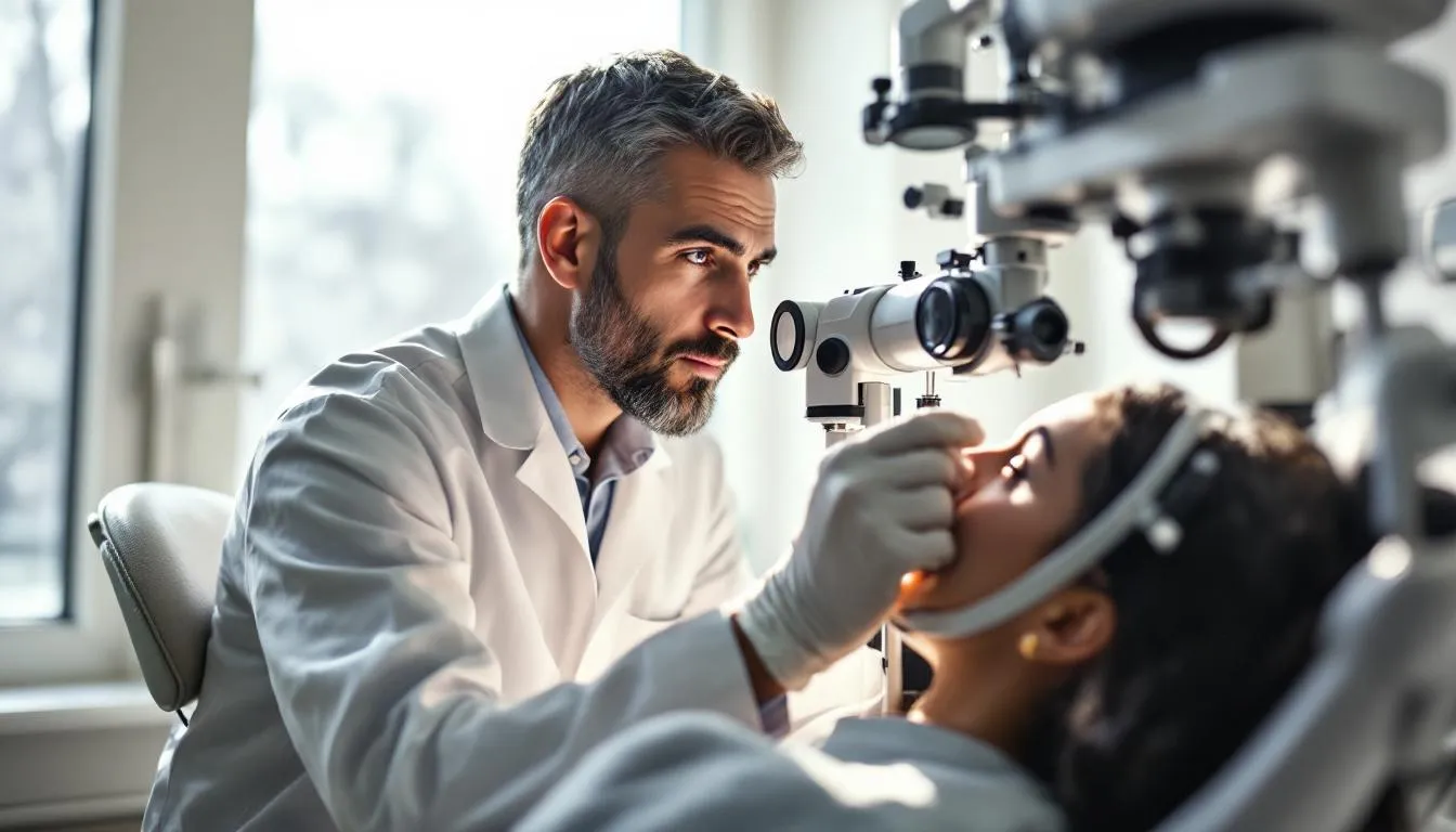
Ophthalmology is the branch of medicine devoted to the diagnosis, treatment, and prevention of diseases and disorders of the eye and visual system. Ophthalmologists are physicians — MDs or DOs — who can prescribe medications, perform surgery, and manage everything from routine vision problems to complex conditions threatening permanent blindness.
The Eye: A Quick Anatomy Lesson
Before you can understand eye diseases, you need a basic map of the eye. And it’s genuinely remarkable engineering.
Light enters through the cornea — the clear, dome-shaped front surface that does about 65-75% of the eye’s focusing. Behind the cornea sits the iris (the colored part) surrounding the pupil, which dilates and constricts to control how much light enters. The lens, just behind the iris, fine-tunes focus by changing shape — a process called accommodation that gradually diminishes with age (which is why nearly everyone needs reading glasses after 40).
Light passes through the vitreous humor — a clear gel filling the main cavity of the eye — and hits the retina, a thin layer of neural tissue lining the back of the eye. The retina contains about 120 million rod cells (for low-light and peripheral vision) and 6-7 million cone cells (for color and detail). These photoreceptors convert light into electrical signals that travel via the optic nerve to the brain’s visual cortex, where the actual “seeing” happens.
The entire structure is about 2.4 centimeters in diameter and weighs roughly 7.5 grams. It can distinguish about 10 million different colors and resolve detail as fine as 0.59 arc minutes — roughly equivalent to spotting a quarter from 75 feet away.
Common Eye Conditions
Ophthalmologists deal with a wide spectrum of conditions, from minor annoyances to sight-threatening emergencies.
Refractive Errors
The most common vision problems are simply issues of focus. Myopia (nearsightedness) means the eye is too long or the cornea too curved — light focuses in front of the retina, making distant objects blurry. Hyperopia (farsightedness) is the opposite. Astigmatism occurs when the cornea has an irregular shape, causing distorted or blurred vision at all distances. Presbyopia — the age-related loss of near focus — hits almost everyone by their mid-40s as the lens stiffens and loses its ability to change shape.
Globally, about 2.7 billion people have uncorrected refractive errors. In wealthy countries, this is mostly a minor inconvenience — glasses or contacts fix it. In low-income countries, lack of access to corrective lenses remains a major cause of functional blindness and lost productivity.
Myopia is also increasing at alarming rates worldwide. In East Asia, up to 90% of young adults in some urban populations are now myopic, compared to 10-30% two generations ago. Research strongly suggests that lack of outdoor time in childhood is a key driver — children who spend more time outside have lower myopia rates, likely due to the brightness of natural light and the need to focus at varying distances.
Cataracts
A cataract is a clouding of the eye’s natural lens. It’s the leading cause of blindness worldwide — about 17 million people are blind from cataracts, mostly in developing countries where surgical access is limited.
In developed countries, cataract surgery is one of the most commonly performed surgical procedures — about 4 million per year in the United States alone. The operation takes about 15-20 minutes: the clouded lens is broken apart by ultrasound (phacoemulsification), removed through a tiny incision, and replaced with a clear artificial lens (intraocular lens, or IOL). Success rates exceed 99%, and patients typically notice dramatically improved vision within a day or two.
Modern IOL technology is impressive. Multifocal and extended-depth-of-focus lenses can reduce or eliminate the need for glasses after surgery. Toric lenses correct astigmatism simultaneously.
Glaucoma
Glaucoma is a group of conditions that damage the optic nerve, usually (but not always) associated with elevated pressure inside the eye. It’s called “the sneak thief of sight” because the most common form — open-angle glaucoma — causes gradual, painless peripheral vision loss that most people don’t notice until significant damage has occurred.
About 80 million people worldwide have glaucoma. It’s the leading cause of irreversible blindness globally. Risk factors include age over 60, African or Hispanic ancestry, family history, elevated eye pressure, and high myopia.
Treatment focuses on lowering intraocular pressure through eye drops, laser procedures (selective laser trabeculoplasty), or surgery (trabeculectomy, tube shunt implantation, or minimally invasive glaucoma surgery — MIGS). None of these reverse existing damage, but they can slow or halt progression. That’s why routine screening is so important — catching glaucoma early means preserving more vision.
Age-Related Macular Degeneration (AMD)
AMD affects the macula — the central part of the retina responsible for sharp, detailed vision. It’s the leading cause of vision loss in people over 50 in developed countries, affecting about 196 million people worldwide.
The “dry” form (about 80% of cases) involves gradual thinning of the macula and accumulation of yellow deposits called drusen. There’s no cure, but nutritional supplements (the AREDS2 formula — vitamins C and E, zinc, copper, lutein, and zeaxanthin) can slow progression in some patients.
The “wet” form (20% of cases) involves abnormal blood vessel growth beneath the retina. These vessels leak fluid and blood, causing rapid, severe vision loss. Treatment with anti-VEGF injections — drugs like ranibizumab (Lucentis), aflibercept (Eylea), and bevacizumab (Avastin) — has been genuinely life-changing for patients with wet AMD. Before these drugs existed (the first was approved in 2004), wet AMD almost always led to severe central vision loss. Now, most patients maintain or improve their vision with regular injections.
Diabetic Eye Disease
Diabetes damages blood vessels throughout the body, and the retina’s tiny vessels are particularly vulnerable. Diabetic retinopathy affects roughly one-third of people with diabetes. In its early stages (non-proliferative), blood vessels leak or develop microaneurysms. In advanced stages (proliferative), new abnormal blood vessels grow on the retina’s surface and can bleed into the vitreous, cause retinal detachment, and lead to blindness.
Diabetic macular edema — swelling of the macula from leaking blood vessels — is the most common cause of vision loss in diabetic patients. Treatment includes anti-VEGF injections, laser photocoagulation, and — most importantly — good blood sugar control. The Diabetes Control and Complications Trial showed that tight glycemic control reduced the risk of diabetic retinopathy progression by 76%.
Surgical Subspecialties
Ophthalmology is one of the most surgery-heavy medical specialties. Common procedures include:
Refractive surgery — LASIK, PRK (photorefractive keratectomy), and SMILE (small incision lenticule extraction) reshape the cornea to correct refractive errors. LASIK has been performed on over 40 million people worldwide, with patient satisfaction rates consistently above 95%.
Retinal surgery — vitrectomy (removing the vitreous gel to access and repair the retina), retinal detachment repair, and macular hole surgery. These are microsurgical procedures performed with instruments that are sometimes less than 0.5 millimeters in diameter.
Corneal transplant — replacing a damaged cornea with donor tissue. About 49,000 corneal transplants are performed in the U.S. annually, making the cornea the most commonly transplanted tissue in the body.
Oculoplastic surgery — procedures on the eyelids, tear ducts, and orbit (the bony socket surrounding the eye). This includes ptosis repair (drooping eyelids), blocked tear duct surgery, and orbital fracture reconstruction.
The Future of the Field
Ophthalmology is at the forefront of some genuinely exciting medical technology. Gene therapy for inherited retinal diseases is already a reality — voretigene neparvovec (Luxturna), approved in 2017, treats a specific form of inherited blindness by delivering a functional copy of the RPE65 gene directly to retinal cells.
Artificial intelligence is being integrated into screening programs — algorithms that analyze retinal photographs can detect diabetic retinopathy with accuracy comparable to trained ophthalmologists, potentially enabling mass screening in areas without eye doctors.
Retinal prosthetics (bionic eyes) remain experimental but have produced some vision restoration in patients with complete blindness. The technology is crude by biological standards — current devices provide very low resolution — but the concept works, and improvements are ongoing.
Stem cell-based therapies for macular degeneration and other retinal diseases are in clinical trials, with early results showing promise for regenerating damaged retinal tissue.
When to See an Ophthalmologist
Some eye symptoms demand urgent attention: sudden vision loss, sudden onset of flashing lights or a shower of new floaters (possible retinal detachment), eye pain with redness and nausea (possible acute angle-closure glaucoma), and sudden onset of a curtain or shadow across your field of vision.
For everything else, regular eye exams are your best defense. Many sight-threatening conditions — glaucoma, diabetic retinopathy, early macular degeneration — have no symptoms in their early stages. The only way to catch them is to look. Given that vision is arguably our most relied-upon sense, periodic professional inspection seems like a reasonable investment.
Frequently Asked Questions
What is the difference between an ophthalmologist and an optometrist?
An ophthalmologist is a medical doctor (MD or DO) who completed medical school, a one-year internship, and a three-year ophthalmology residency. They can diagnose and treat all eye diseases, prescribe medications, and perform surgery. An optometrist (OD) completes a four-year Doctor of Optometry program after college and focuses on vision testing, prescribing glasses and contacts, and diagnosing common eye conditions. Optometrists refer complex cases and surgical needs to ophthalmologists.
How often should you get an eye exam?
The American Academy of Ophthalmology recommends a baseline thorough eye exam at age 40, when early signs of disease or vision changes may appear. Before 40, adults with no risk factors should have exams every 5-10 years. After 40, every 2-4 years. After 55, every 1-3 years. After 65, every 1-2 years. People with diabetes, a family history of glaucoma, or other risk factors should be examined more frequently — typically annually.
What is LASIK surgery and who is a good candidate?
LASIK (Laser-Assisted in Situ Keratomileusis) is a refractive surgery that reshapes the cornea to correct nearsightedness, farsightedness, and astigmatism. A laser creates a thin flap in the cornea, reshapes the underlying tissue, then repositions the flap. Good candidates are generally 18 or older, have had a stable prescription for at least a year, have healthy corneas of adequate thickness, and don't have certain conditions like keratoconus or severe dry eye. About 96% of patients achieve 20/20 vision or better.
Can glaucoma be cured?
No, glaucoma cannot be cured, but it can be managed. The damage to the optic nerve is permanent and irreversible, but treatment — eye drops, laser procedures, or surgery — can lower eye pressure and slow or stop further damage. That's why early detection is critical: by the time you notice vision loss from glaucoma, significant irreversible damage has already occurred. Regular eye exams are the only way to catch it early.
Further Reading
Cite this article
APA
WhatIs.site. (2025). What Is Ophthalmology?. Retrieved May 27, 2026, from https://whatis.site/ophthalmology MLA
"What Is Ophthalmology?." WhatIs.site, July 15, 2025, https://whatis.site/ophthalmology. Accessed May 27, 2026. Chicago
WhatIs.site. "What Is Ophthalmology?." Last modified May 12, 2026. https://whatis.site/ophthalmology. HTML
<a href="https://whatis.site/ophthalmology">What Is Ophthalmology?</a> — WhatIs.site Related Articles
What Is Optometry?
Optometry is the healthcare profession focused on vision care and eye health. Learn about eye exams, corrective lenses, common conditions, and careers.
scienceWhat Is Anatomy?
Anatomy is the study of body structure in living organisms. Learn about gross and microscopic anatomy, organ systems, history, and why it matters in medicine.
health amp wellnessWhat Is Nursing?
Nursing is a healthcare profession focused on patient care, health promotion, and illness prevention. Learn about nursing roles, education, and specialties.