Table of Contents
What Is Comparative Anatomy?
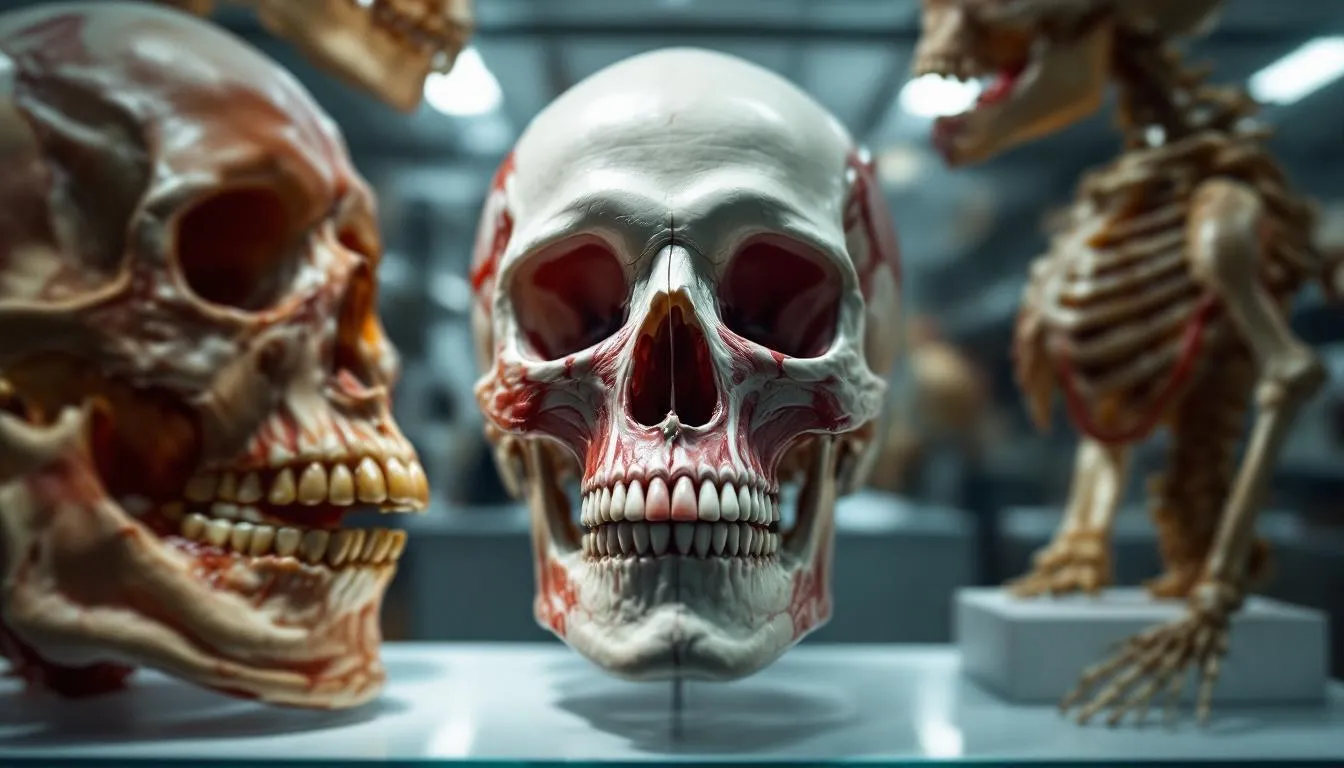
Comparative anatomy is the branch of biology that studies the similarities and differences in the physical structures of different species to understand evolutionary relationships, functional adaptations, and the principles that govern body design across the animal kingdom. By comparing how bones, organs, and tissues are arranged in different organisms, scientists can reconstruct evolutionary history, understand how structures adapt to different environments, and predict how organisms will respond to new challenges.
The Insight That Changed Biology
Here’s the observation that launched comparative anatomy as a serious science: your arm and a whale’s flipper contain the same bones.
Look at your arm. You’ve got one bone in the upper arm (humerus), two in the forearm (radius and ulna), a cluster of small wrist bones (carpals), and five sets of finger bones (metacarpals and phalanges). Now look at a whale’s flipper. Same bones. Same arrangement. Humerus, radius, ulna, carpals, metacarpals, phalanges — they’re shortened, flattened, and modified for swimming, but they’re the same bones in the same relative positions.
Now look at a bat’s wing. Same bones again — but the finger bones are enormously elongated to support the wing membrane. A horse’s leg? Same bones — but the horse walks on a single modified finger (the third digit), with the other digits reduced to tiny splints.
This pattern repeats across every tetrapod (four-limbed vertebrate) on Earth. Frogs, lizards, birds, cats, humans — all share the same fundamental limb bone arrangement. The bones are stretched, compressed, fused, or reduced in various ways, but the underlying pattern is unmistakable.
Before Darwin, this pattern was puzzling. If each species was independently designed for its specific lifestyle, why use the same bones in such radically different ways? Why give a whale the same arm bones as a human when a completely different flipper design might work better? The answer, as Darwin recognized, is descent with modification. All tetrapods inherited this limb plan from a common ancestor, and evolution modified it for different functions in different lineages.
A Brief History
Comparative anatomy has ancient roots. Aristotle dissected animals and classified them based on structural similarities over 2,300 years ago. His Historia Animalium organized hundreds of species based on observable anatomical features — an impressive achievement for someone working without microscopes, X-rays, or any concept of evolution.
But the field truly came into its own in the 18th and 19th centuries. Georges Cuvier (1769-1832) is often called the father of comparative anatomy. Working at the National Museum of Natural History in Paris, Cuvier established the principle of “correlation of parts” — the idea that an organism’s body parts are functionally interdependent. If you find a skull with sharp teeth, you can predict clawed feet and a short digestive tract (characteristics of a carnivore). The whole body hangs together as a functional unit.
Cuvier’s principle was so powerful that he could reconstruct extinct animals from fragmentary fossils. Hand him a few bones, and he could tell you what kind of animal it was, how it moved, and what it ate. This made paleontology possible as a science — you can’t dig up a complete dinosaur very often, but you can usually find enough bones for comparative analysis.
Richard Owen, working in London in the mid-1800s, coined the term “homology” to describe structural similarities between species. Owen noticed the consistent pattern in vertebrate limbs but — crucially — interpreted it differently than Darwin would. Owen believed homologies reflected a divine “archetype,” a perfect body plan from which all vertebrates were variations. Darwin reinterpreted the same evidence as descent from a common ancestor. Same observations, radically different explanations.
Homology: The Central Concept
Homologous structures are the bread and butter of comparative anatomy. Two structures are homologous if they share a common evolutionary origin, regardless of their current function.
The vertebrate limb is the most famous example, but homologies are everywhere.
The mammalian ear ossicles: Your middle ear contains three tiny bones — the malleus, incus, and stapes — that transmit sound from your eardrum to your inner ear. Here’s the wild part: two of these bones (the malleus and incus) evolved from jaw bones that reptiles use for eating. In the fossil record, you can actually trace the gradual migration of these bones from the jaw to the ear across millions of years of mammalian evolution. Reptilian jaw bones became mammalian hearing apparatus. Same bones, completely different function.
The recurrent laryngeal nerve: In all mammals, this nerve travels from the brain down into the chest, loops around the aorta, and then back up to the larynx (voice box). In a human, the detour adds a few inches. In a giraffe, the nerve travels about 15 feet to reach an organ only inches away from its starting point. This absurd routing makes no sense from a design perspective but perfect sense evolutionarily — the nerve’s path was established in fish ancestors (where it took a direct route to gill structures), and as the neck elongated in mammalian evolution, the nerve was stretched along with it.
Pentadactyl limbs: The five-fingered (pentadactyl) plan appears across virtually all tetrapods. Some species have reduced the number — horses have one functional digit, birds have fused and reduced digits — but the developmental blueprint starts with five in almost all cases. This consistency across hundreds of millions of years and millions of species is powerful evidence of common descent.
Analogy: When Evolution Converges
Not all similarities between species indicate shared ancestry. Sometimes, unrelated organisms evolve similar structures independently because they face similar environmental challenges. These are analogous structures — similar in function but different in origin.
Wings provide the textbook example. Bird wings, bat wings, insect wings, and pterosaur wings all serve the same function (flight), but they evolved completely independently. Bird wings are modified forelimbs with feathers. Bat wings are modified hands with membrane stretched between elongated fingers. Insect wings are extensions of the exoskeleton with no skeletal support at all. Pterosaur wings were supported by a single elongated fourth finger.
Camera eyes evolved independently in vertebrates and cephalopods (octopuses and squid). Both have a lens that focuses light onto a retina, but the detailed structure differs — most strikingly, the vertebrate retina is “inside out” (the nerve fibers are on the front surface, creating a blind spot), while the cephalopod retina has the nerve fibers behind the photoreceptors (no blind spot). Same problem, similar solution, different engineering.
Streamlined body shapes appear in dolphins (mammals), sharks (fish), and ichthyosaurs (extinct reptiles). These three lineages are only distantly related, but they all evolved torpedo-shaped bodies for moving efficiently through water. The physics of fluid dynamics imposed similar body shapes on completely different starting points.
This phenomenon — convergent evolution — is actually powerful evidence for evolution. It shows that natural selection predictably produces similar solutions to similar problems. And comparative anatomy is how we distinguish convergence from common ancestry: by looking past superficial similarity to examine underlying structural details.
Vestigial Structures: Evolution’s Leftovers
Some structures that made perfect sense in ancestral species persist in modified form long after they’ve lost their original function. These vestigial structures are among comparative anatomy’s most compelling evidence for evolution.
The human appendix is a reduced remnant of the cecum, a much larger organ in herbivorous mammals that helps ferment plant material. Our primate ancestors shifted toward a more omnivorous diet, and the cecum shrank. The appendix may retain minor immune functions, but it’s a shadow of its ancestral organ.
Whale pelvic bones are small, free-floating bones buried in muscle tissue — remnants of the hind limbs their ancestors used to walk on land. Some whale species occasionally produce individuals with tiny, visible hind limbs, showing that the genetic instructions for leg development are still present but usually suppressed.
The human coccyx (tailbone) is the remnant of a tail that our primate ancestors lost millions of years ago. It acts as an attachment point for some pelvic muscles, but it’s a vestige of a much more substantial structure.
Flightless bird wings — ostriches, emus, kiwis — retain wings that are far too small for flight. These wings are homologous to the fully functional wings of flying birds, reduced through evolution as these species adapted to ground-dwelling lifestyles.
Python pelvic spurs: Pythons and boas have tiny claw-like structures near their tails — remnants of hind legs from their lizard ancestors. These spurs have been repurposed for gripping during mating but are clearly vestigial limbs.
Embryology and Comparative Development
Comparative anatomy extends beyond adult structures to embryonic development, and this is where some of the most striking patterns emerge.
Karl Ernst von Baer noticed in the 1820s that embryos of different vertebrate species look remarkably similar in their early stages. A four-week human embryo has pharyngeal arches (structures that become gills in fish but develop into jaw, ear, and throat structures in mammals), a tail, and a body plan that’s nearly indistinguishable from a chicken or pig embryo at the same stage.
As development proceeds, species-specific features emerge. The pharyngeal arches develop into gills in fish but into the jaw, hyoid bone, and larynx in humans. The tail is absorbed in humans but elongates in mice and cats. The limb buds that look identical in early embryos develop into arms, wings, flippers, or legs depending on the species.
This pattern — similar early development diverging into different adult forms — reflects shared developmental genes. The same Hox genes that control body segmentation in fruit flies also control body segmentation in humans. The same signaling molecules (sonic hedgehog, bone morphogenetic proteins) pattern limbs in all tetrapods. Evolution modifies existing developmental programs rather than building new ones from scratch.
Frankly, the molecular evidence for shared developmental programs has been one of the most powerful confirmations of what comparative anatomists suspected from structure alone. The same toolkit genes, modified in different ways, produce the stunning variety of animal body plans we see across the animal kingdom.
Functional Morphology: Form Follows Function
Comparative anatomy isn’t just about evolutionary history — it’s also about understanding how structures work. Functional morphology studies the relationship between an organism’s structure and how it uses that structure.
Skull and jaw mechanics: Carnivore skulls have large sagittal crests (ridges on top for jaw muscle attachment), sharp shearing teeth, and jaw joints that allow powerful vertical biting but minimal side-to-side movement. Herbivore skulls have flatter surfaces, broad grinding teeth, and jaw joints that permit extensive lateral movement for chewing tough plant material. The skull’s architecture directly reflects diet.
Limb proportions and locomotion: Animals that run fast have long, thin limbs with reduced digits. Animals that dig have short, powerful limbs with large claws. Animals that swing through trees have long, flexible arms with grasping hands. Animals that swim have flattened, paddle-like limbs. You can predict an animal’s lifestyle from its limb proportions — which is exactly what paleontologists do with fossil species.
Respiratory systems: Bird lungs use a one-way flow system with air sacs, achieving far more efficient gas exchange than mammalian tidal-flow lungs. This efficiency is what allows birds to fly at extreme altitudes — bar-headed geese migrate over the Himalayas at 29,000 feet, where oxygen levels are about one-third of sea level. Comparing bird and mammalian respiratory anatomy reveals how different engineering solutions address the same fundamental problem of oxygen delivery.
Modern Comparative Anatomy
The field has been transformed by technology and by integration with molecular biology, but its core methods remain essential.
CT scanning allows researchers to examine internal anatomy without dissection, creating detailed 3D models of skeletal and soft tissue structures. Micro-CT can image structures at resolutions below one micrometer, revealing details invisible to the naked eye.
3D morphometrics uses statistical analysis of landmark positions to quantify shape differences between species, populations, and individuals. This allows precise, numerical comparisons that go beyond subjective visual assessment.
Phylogenetic comparative methods combine anatomical data with molecular phylogenies (evolutionary trees based on DNA) to test hypotheses about how structures evolved. Did a particular structure evolve once or multiple times? Did body size increase or decrease along a particular evolutionary lineage? These questions require both anatomical and molecular data.
Evo-devo (evolutionary developmental biology) bridges comparative anatomy and genetics by studying how changes in developmental genes produce changes in body structure. A mutation in a single Hox gene can transform one body segment’s structures into another’s — demonstrating that the connection between genes and anatomy is direct and mechanistic.
Why It Still Matters
With genomics and molecular biology, you might wonder whether comparing bones and organs is still relevant. It absolutely is, for several reasons.
Fossils preserve anatomy, not DNA. For the vast majority of evolutionary history — the 3.5 billion years before the last few hundred thousand — anatomical comparison is the primary way to reconstruct evolutionary relationships. You can sequence a mammoth’s genome, but you can’t sequence a dinosaur’s.
Functional questions require anatomical answers. Understanding how an animal moves, feeds, senses, and reproduces requires knowing how its body is built. Genomics tells you what genes are present; comparative anatomy tells you what those genes actually built.
Medical research depends on anatomical comparison. When researchers use mice, rats, or monkeys as models for human disease, they’re making comparative anatomical assumptions — that these species’ organs are similar enough to humans’ that findings will transfer. Understanding exactly where those similarities hold and where they break down is a comparative anatomy question.
Conservation biology uses comparative anatomy to assess species’ vulnerability to environmental change. Species with specialized anatomical adaptations (narrow dietary requirements, limited locomotor abilities) are generally more vulnerable to habitat disruption than species with more generalized anatomy.
Comparative anatomy began with the simple observation that different animals share the same bones. From that observation grew an entire framework for understanding life’s diversity, history, and interconnectedness. The bones are still telling us stories — we just need to know how to read them.
Frequently Asked Questions
What is the difference between homologous and analogous structures?
Homologous structures share a common evolutionary origin but may serve different functions — like a human arm, a whale flipper, and a bat wing, which all derive from the same ancestral limb bones. Analogous structures serve similar functions but evolved independently — like bird wings and butterfly wings, which both enable flight but have completely different underlying structures.
How did comparative anatomy help prove evolution?
Comparative anatomy provided some of the earliest and most compelling evidence for evolution. The consistent pattern of homologous structures across diverse species — the same bones rearranged for different functions — is most logically explained by descent from a common ancestor, not by independent creation of each species.
What are vestigial structures?
Vestigial structures are body parts that have lost most or all of their original function through evolution. Examples include the human appendix (reduced from a larger cecum in plant-eating ancestors), wisdom teeth (useful for our ancestors' tougher diet but problematic for modern jaw sizes), and the pelvic bones in whales (remnants from their four-legged terrestrial ancestors).
Is comparative anatomy still relevant today?
Absolutely. While molecular genetics has transformed evolutionary biology, comparative anatomy remains essential for interpreting fossil records (which preserve anatomy but rarely DNA), understanding functional morphology, developing animal models for medical research, and studying how physical structures constrain or enable behavioral adaptations.
Further Reading
Cite this article
APA
WhatIs.site. (2026). What Is Comparative Anatomy?. Retrieved May 27, 2026, from https://whatis.site/comparative-anatomy MLA
"What Is Comparative Anatomy?." WhatIs.site, March 6, 2026, https://whatis.site/comparative-anatomy. Accessed May 27, 2026. Chicago
WhatIs.site. "What Is Comparative Anatomy?." Last modified May 12, 2026. https://whatis.site/comparative-anatomy. HTML
<a href="https://whatis.site/comparative-anatomy">What Is Comparative Anatomy?</a> — WhatIs.site Related Articles
What Is Anatomy?
Anatomy is the study of body structure in living organisms. Learn about gross and microscopic anatomy, organ systems, history, and why it matters in medicine.
scienceWhat Is Animal Behavior?
Animal behavior is the scientific study of how animals act, interact, and respond to their environment — covering instinct, learning, and communication.
scienceWhat Is Anthropology?
Anthropology is the study of humans—past and present—across cultures, biology, language, and societies. Learn its branches, methods, and why it matters.
scienceWhat Is Agriculture?
Agriculture is the practice of cultivating plants, raising animals, and processing natural resources to produce food, fiber, and fuel.
scienceWhat Is Acoustics?
Acoustics is the science of sound: how it's produced, transmitted, and received. Learn about sound waves, architectural acoustics, noise control, and more.